


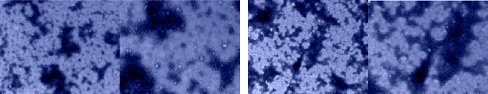
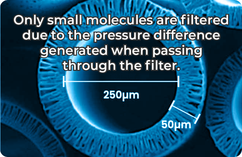
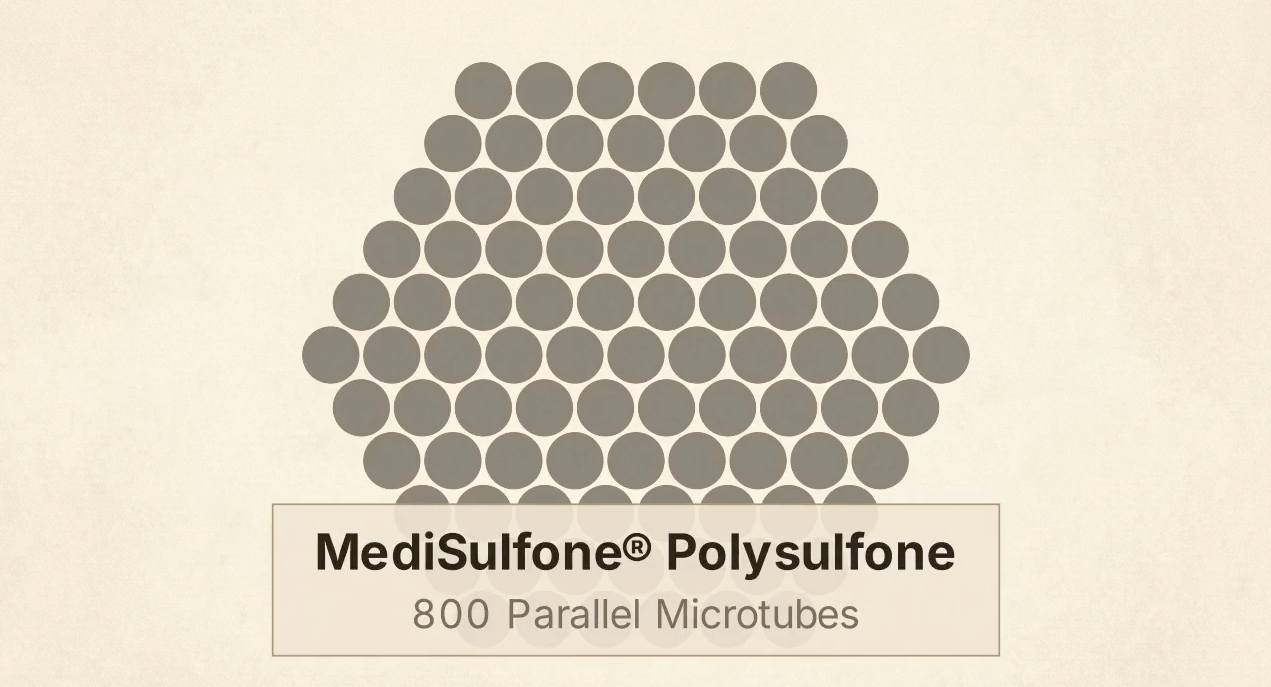
Analytical testing measured approximately 30 trillion particles per milliliter. Flow cytometry confirmed expression of CD9 (93.2%), CD63 (90.1%), and CD81 (69.1%) — the three canonical EV markers recognized by the MISEV2023 international standard. TEM confirmed vesicular morphology. All EVs (30–1,000 nm) are physically retained by the membrane’s 5 nm pores.
Source: StemExoOne Co., Ltd., 2024 (commissioned analytical testing).

The 15 kDa membrane retains all plasma proteins above its molecular weight cutoff: fibrinogen (340 kDa) for fibrin scaffold formation, alpha-2-macroglobulin (720 kDa) for protease inhibition, fibronectin (440 kDa) for cell adhesion, and albumin (66.5 kDa) for growth factor transport. Published studies report approximately 2× protein enrichment using ultrafiltration (Mazzucco et al. 2024, Frontiers in Bioengineering and Biotechnology).

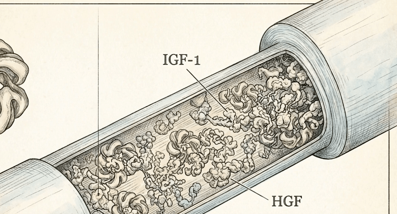
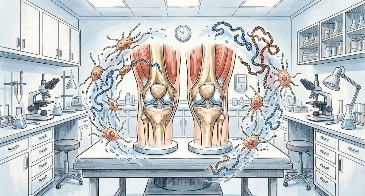
Platelet-derived growth factors (PDGF, VEGF, TGF-β, EGF) are concentrated alongside extraplatelet factors (IGF-1, HGF) that standard centrifugation PRP cannot enrich. Published data show IGF-1 is the only growth factor correlated with cell proliferation, and it resides in plasma rather than platelets (Beitia et al. 2023, International Journal of Molecular Sciences).

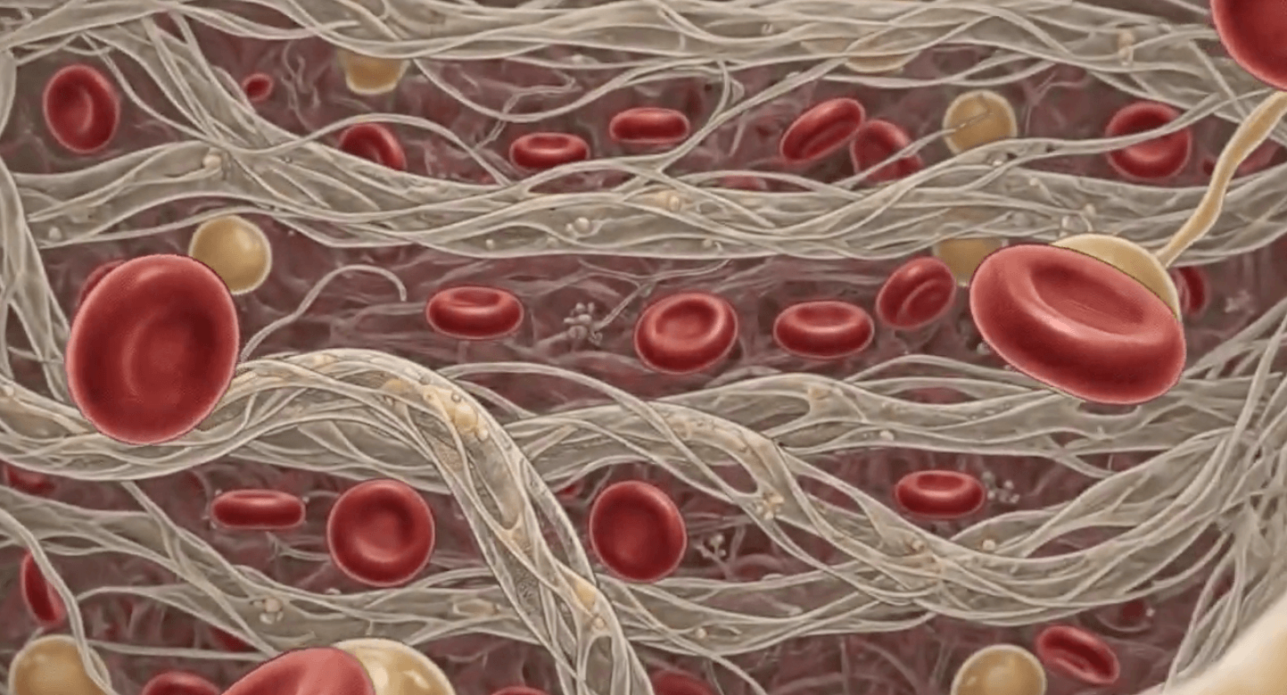
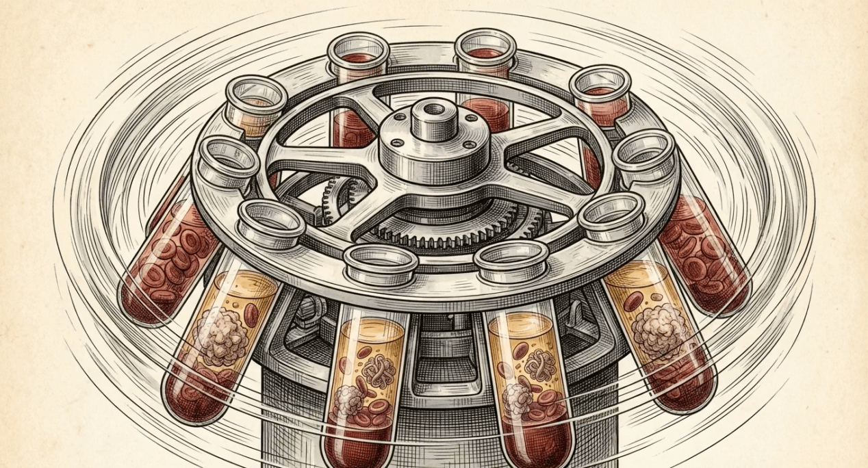
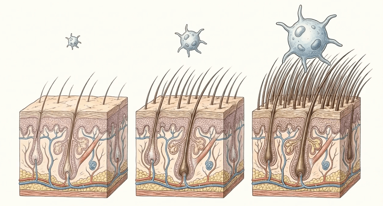
Platelets are retained above the gel barrier during centrifugation and concentrated during ultrafiltration. The larger output volume (approximately 6–7 mL) is designed to deliver a higher total platelet dose to the treatment site.

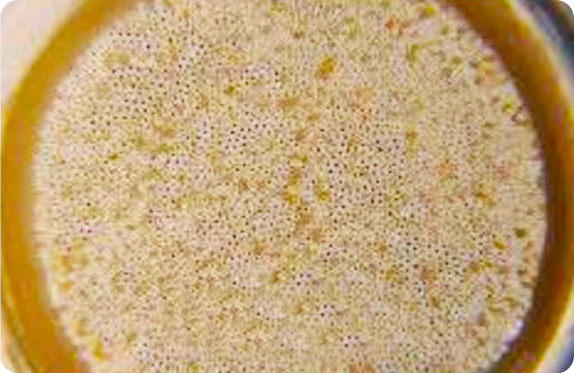
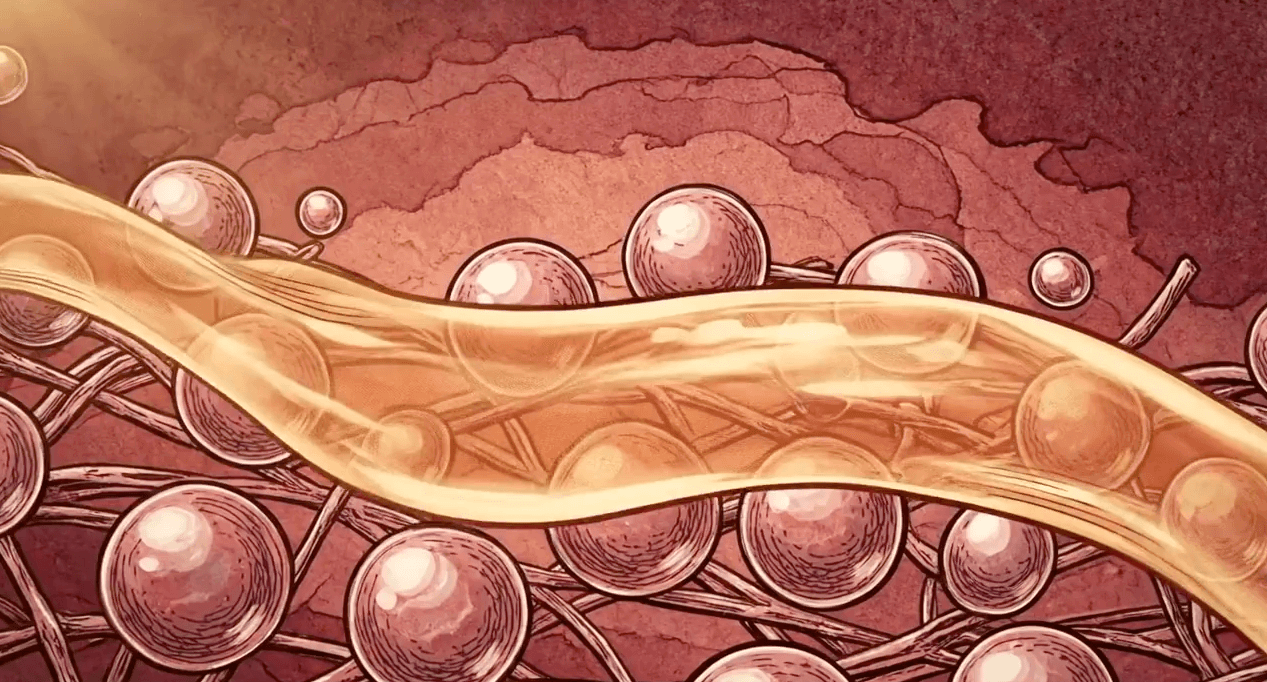
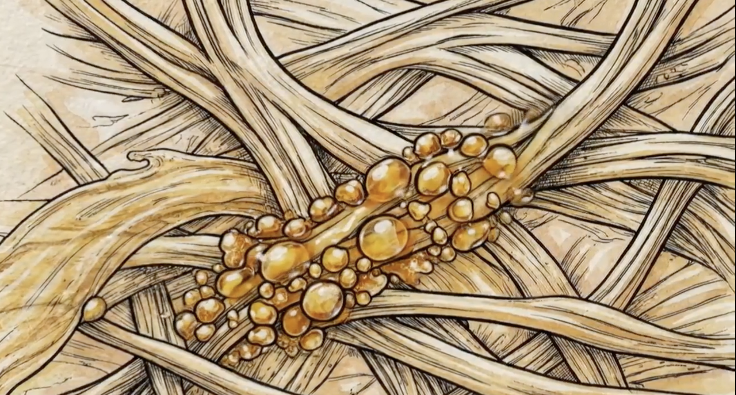
Concentrated fibrinogen, upon activation, forms a dense autologous fibrin matrix. Published research shows fibrinogen binds at least 15 growth factors with nanomolar affinity, creating a sustained-release reservoir lasting up to 7 days (Martino et al. 2013, PNAS).


IGF-1

HGF

Alpha-2-macroglobulin (A2M)

Extracellular vesicles


PLATELET AND EV SIGNALING
GROWTH FACTOR COOPERATION

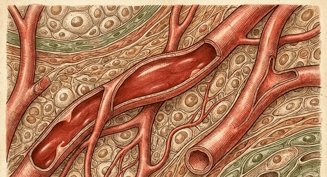
INFLAMMATION MODULATION

SCAFFOLD FORMATION

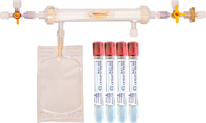

CE-MARKED COMPONENTS

100% AUTOLOGOUS

CHARACTERIZED EXOSOMES

DUAL CONCENTRATION
REPRODUCIBLE

DEFINED PARAMETERS
